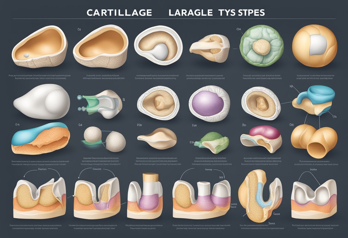
Types Of Cartilage
Cartilage is a type of connective tissue that is present in various parts of the human body, including joints, the rib cage, intervertebral discs, the ear, and the nose. It is a semi-rigid but flexible tissue that provides support, shock absorption, and bone repair. Cartilage is composed of chondrocytes, which are the main cell type in cartilage, and a ground substance called chondroitin sulfate.
There are three types of cartilage in the human body: hyaline, fibrous, and elastic. Hyaline cartilage is the most widespread type and is found in the trachea, bronchi, and at the ends of long bones. Fibrous cartilage is found in the intervertebral discs and the pubic symphysis, while elastic cartilage is found in the ear and the epiglottis. Each type of cartilage has its own unique properties and functions.
Understanding the different types of cartilage is important for maintaining joint health and preventing conditions such as osteoarthritis and rheumatoid arthritis. It is also important for researchers who are studying cartilage growth and repair, as well as the biomechanical properties of cartilage. By gaining a deeper understanding of cartilage, we can develop new treatments and therapies to improve joint health and overall quality of life.
Key Takeaways
- Cartilage is a type of connective tissue that is semi-rigid but flexible and provides support, shock absorption, and bone repair.
- There are three types of cartilage in the human body: hyaline, fibrous, and elastic, each with its own unique properties and functions.
- Understanding the different types of cartilage is important for maintaining joint health, preventing conditions such as osteoarthritis and rheumatoid arthritis, and developing new treatments and therapies.
Basic Structure and Function of Cartilage

Cartilage is a type of connective tissue that is found in various areas of the body, such as the joints, ear, nose, and airway. It is composed of chondrocytes, which are specialized cells that produce and maintain the cartilage matrix. The matrix is made up of collagen fibers and proteoglycans, which give cartilage its strength and elasticity.
Components of Cartilage Matrix
Collagen is the most abundant protein in the cartilage matrix. It provides tensile strength and helps to resist stretching and tearing. Proteoglycans are large molecules that are composed of a protein core and long chains of sugar molecules called glycosaminoglycans. These molecules attract water, which gives cartilage its ability to resist compression and absorb shock.
Cartilage Types and Locations
There are three main types of cartilage: hyaline, elastic, and fibrocartilage. Hyaline cartilage is the most common type of cartilage and is found in the nose, trachea, larynx, and joints. Elastic cartilage is found in the ear and epiglottis. Fibrocartilage is found in the intervertebral discs, pubic symphysis, and the insertion points of tendons and ligaments.
Hyaline cartilage is the most abundant type of cartilage and is characterized by its glassy appearance. It provides a smooth surface for joint movement and helps to distribute weight evenly across the joint. Elastic cartilage contains more elastic fibers than hyaline cartilage and is more flexible. It provides support and maintains the shape of structures such as the ear and nose. Fibrocartilage is the strongest type of cartilage and is characterized by its high collagen content. It is found in areas that are subject to high stress and helps to absorb shock.
In summary, cartilage is a specialized type of connective tissue that provides support, cushioning, and flexibility to various structures in the body. Its composition and structure vary depending on its location and function.
Cartilage Growth and Repair
Cartilage is a specialized connective tissue that has a limited capacity for self-repair. However, it can repair itself through a process called chondrogenesis, which is the formation of cartilage from mesenchymal cells.
Chondrogenesis in Embryo
During embryonic development, mesenchyme cells differentiate into chondroblasts, which produce the extracellular matrix of cartilage. Chondrocytes, which are mature chondroblasts, maintain the extracellular matrix and are responsible for the growth and maintenance of cartilage.
Chondrogenesis is regulated by various signaling pathways, including the bone morphogenetic protein (BMP), fibroblast growth factor (FGF), and transforming growth factor-beta (TGF-β) pathways. These pathways play a critical role in the differentiation and proliferation of chondrocytes.
Cartilage Healing and Regeneration
Cartilage repair is a slow process due to its avascular nature, which limits the delivery of nutrients and oxygen to the cells. The perichondrium, a layer of connective tissue that surrounds cartilage, can provide a source of chondrogenic cells for cartilage repair.
The extracellular matrix of cartilage is composed of collagen, proteoglycans, and glycosaminoglycans (GAGs), including chondroitin sulfate. These components are essential for the mechanical properties of cartilage, including its ability to withstand compressive forces.
Cartilage healing and regeneration can occur through two processes: fibrocartilage repair and hyaline cartilage repair. Fibrocartilage repair involves the formation of fibrous tissue, which is less durable than hyaline cartilage. Hyaline cartilage repair involves the production of new cartilage tissue, which is more durable than fibrous tissue.
In conclusion, cartilage growth and repair are complex processes that involve the differentiation and proliferation of chondrocytes, the extracellular matrix, and various signaling pathways. While cartilage has a limited capacity for self-repair, it can repair itself through chondrogenesis and the perichondrium. The extracellular matrix of cartilage is critical for its mechanical properties, and cartilage repair can occur through fibrocartilage or hyaline cartilage repair.
Cartilage in Joint Health and Disease
Cartilage plays a vital role in joint health and disease. It is a strong, flexible connective tissue that protects the joints and bones, acting as a shock absorber throughout the body. There are three types of cartilage in the human body: hyaline cartilage, elastic cartilage, and fibrocartilage.
Role in Movement and Load Distribution
Articular cartilage is the type of cartilage found in joints. It is hyaline cartilage and is responsible for smooth movement and load distribution in joints. Articular cartilage is composed of a dense extracellular matrix (ECM) with a sparse distribution of highly specialized cells called chondrocytes.
Cartilage is essential in the body’s movement as it allows bones to glide over each other. It also helps distribute the load and pressure throughout the joint, preventing damage to the bones. Injuries to cartilage can lead to pain, inflammation, and arthritis.
Degenerative Conditions Affecting Cartilage
Osteoarthritis is a degenerative joint disease that affects articular cartilage. It is a common condition that results from the wear and tear of the cartilage over time. The cartilage becomes thin and damaged, leading to pain, stiffness, and inflammation.
Arthritis is a general term for joint inflammation. It can affect any joint in the body, including those with cartilage. Arthritis can cause pain, stiffness, and swelling in the joints, making it difficult to move.
Subchondral bone is the layer of bone just beneath the articular cartilage. Injuries to the subchondral bone can lead to cartilage damage and degeneration. Spinal disc herniation is a condition that affects the intervertebral discs, which act as shock absorbers between the vertebrae. Injuries to the discs can lead to pain and inflammation.
In conclusion, cartilage plays a crucial role in joint health and disease. It is responsible for smooth movement and load distribution in joints, preventing damage to the bones. Degenerative conditions such as osteoarthritis and arthritis can affect cartilage, leading to pain, stiffness, and inflammation. Injuries to the subchondral bone and intervertebral discs can also cause cartilage damage and degeneration.
Biomechanical Properties of Cartilage
Cartilage is a viscoelastic connective tissue that plays a critical role in the biomechanics of the human body. It is made up of chondrocytes, fibrous tissue, and extracellular matrix (ECM) that contains collagens and proteoglycans. The biomechanical properties of cartilage enable it to resist compressive forces, enhance bone resilience, and provide support on bony areas where there is a need for flexibility.
Resistance to Compressive Forces
Cartilage is highly resistant to compressive forces. It is able to withstand the weight of the body and distribute it evenly across the joint surface. This is due to the high concentration of proteoglycans in the ECM, which attract water molecules and create a hydrostatic pressure that resists compression. The type II collagen fibers in the ECM also help to resist compressive forces.
Elasticity and Tensile Strength
Cartilage is also elastic and has tensile strength. This allows it to deform under load and return to its original shape when the load is removed. The elastin fibers in the ECM give cartilage its elasticity, while the type II collagen fibers provide tensile strength.
Cartilage is avascular, which means it does not have a blood supply. It relies on diffusion of nutrients and waste products from the synovial fluid to maintain its health. Cartilage is also innervated by nerves that transmit pain signals when it is damaged.
Cartilage is surrounded by a joint capsule and is attached to bones by tendons and ligaments. It is a flexible and resilient tissue that provides cushioning and shock absorption to joints, allowing for smooth and pain-free movement.
In summary, cartilage is a biomechanically important tissue that is able to resist compressive forces, provide elasticity and tensile strength, and cushion and protect joints. Its unique composition of fibrous tissue, type II collagen, elastin, and glycosaminoglycans allows it to perform these functions efficiently and effectively.
Advanced Topics in Cartilage Research
Cartilage Tissue Engineering
Cartilage tissue engineering is a rapidly evolving field that aims to develop new methods for repairing and regenerating damaged cartilage. This is particularly important because cartilage has limited capacity for self-repair. Researchers are exploring various strategies to develop new cartilage tissues, including the use of stem cells, scaffolds, and growth factors. The ultimate goal is to develop a tissue-engineered cartilage that can be used to replace damaged or diseased tissue.
One promising approach for cartilage tissue engineering is the use of mesenchymal stem cells (MSCs). These cells have the ability to differentiate into chondrocytes, the cells that produce cartilage. Researchers are also exploring the use of scaffolds to support the growth and development of new cartilage tissue. These scaffolds can be made from a variety of materials, including synthetic polymers and natural substances like collagen and hyaluronic acid.
Another important area of research in cartilage tissue engineering is the use of growth factors and cytokines. These molecules can help to stimulate the growth and differentiation of chondrocytes, leading to the development of new cartilage tissue. Researchers are also exploring the use of fibroblasts, which are cells that produce the extracellular matrix of cartilage, as a potential source of new tissue.
Future Therapies for Cartilage-Related Disorders
In addition to tissue engineering, there are many other potential therapies for cartilage-related disorders. One area of research is the use of lubricin, a glycoprotein that is found in synovial fluid and helps to reduce friction between joint surfaces. Researchers are exploring the use of lubricin as a potential treatment for osteoarthritis, a degenerative joint disease that is characterized by the breakdown of cartilage.
Another area of research is the use of glucosamine sulfate, a dietary supplement that is commonly used to treat osteoarthritis. Glucosamine sulfate is thought to help stimulate the production of new cartilage tissue and may also help to reduce inflammation in the joints.
Researchers are also exploring the microarchitecture of cartilage and how it can be manipulated to promote healing and remodeling. For example, researchers are exploring the use of mechanical loading to stimulate the growth and development of new cartilage tissue. They are also exploring the use of new imaging techniques to better understand the structure and function of cartilage in the body.
Overall, there is still much to learn about cartilage and its role in the body. However, advances in research are providing new insights into the structure and function of cartilage, and are helping to develop new therapies for cartilage-related disorders.


